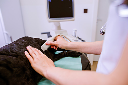
Ultrasound is the second most popular imaging modality in veterinary medicine. Multiple clinical studies have proven that ultrasonography has clear advantages over radiography when diagnosing abdominal organ pathology, non-abdominal soft-tissue conditions, fluid build-up, heart disease, and countless others. Ultrasound is also used in non-diagnostic applications such as safely guiding needles for cystocentesis and cytology and tissue samples.
Ultrasound enables DVMs to see inside an animal’s body without the risks associated with other imaging modalities. Although in human medicine ultrasound is primarily used for pregnancy diagnosis, it has many, many other uses in veterinary medicine.
The following are common indications why a DVM would administer an ultrasound exam:
- Unexplained weight loss
- Persistent diarrhea or vomiting
- Pregnancy
- Fluid build-up in the abdomen
- Abnormal blood-work results
- Chronic infections
- Abnormal urinary habits
- Cardiomegaly on radiography
- Suspected heart failure
- Ligament or tendon tears
- Evaluate the animal before surgery
- Evaluate geriatric patients’ baseline health
When an animal is evaluated with an Ultrasound, the pet will need to be shaved in certain areas where the probe will be used. Ultrasound does not travel through air. Hair traps air and disrupts sound waves which affects the clarity of ultrasound images. Shaving the animal and using a gel-coupling medium improves contact between the patient’s skin and the ultrasound probe, which enhances the quality of images.
In addition, because we do need the pet to stay very still while performing an Ultrasound, the need for light sedation of the pet during the procedure may be required as well. For these reasons, an Ultrasound appointment is a much longer appointment than the typical wellness or even illness appointment.

